EXPLORE · IDENTIFY · UNDERSTAND
Human Atlas: a free interactive 3D anatomy explorer
Human Atlas is a free interactive anatomy explorer that lets you rotate a body, reveal its systems, and inspect individual structures. Start with the model above, then use this guide to make each view more useful.
With the interactive model, you can move between a whole body and an isolated organ without installing an application or creating an account. The adult male reference includes 2,234 selectable model pieces, organized into 15 systems and connected to 3,432 named concepts.
Open the 3D explorer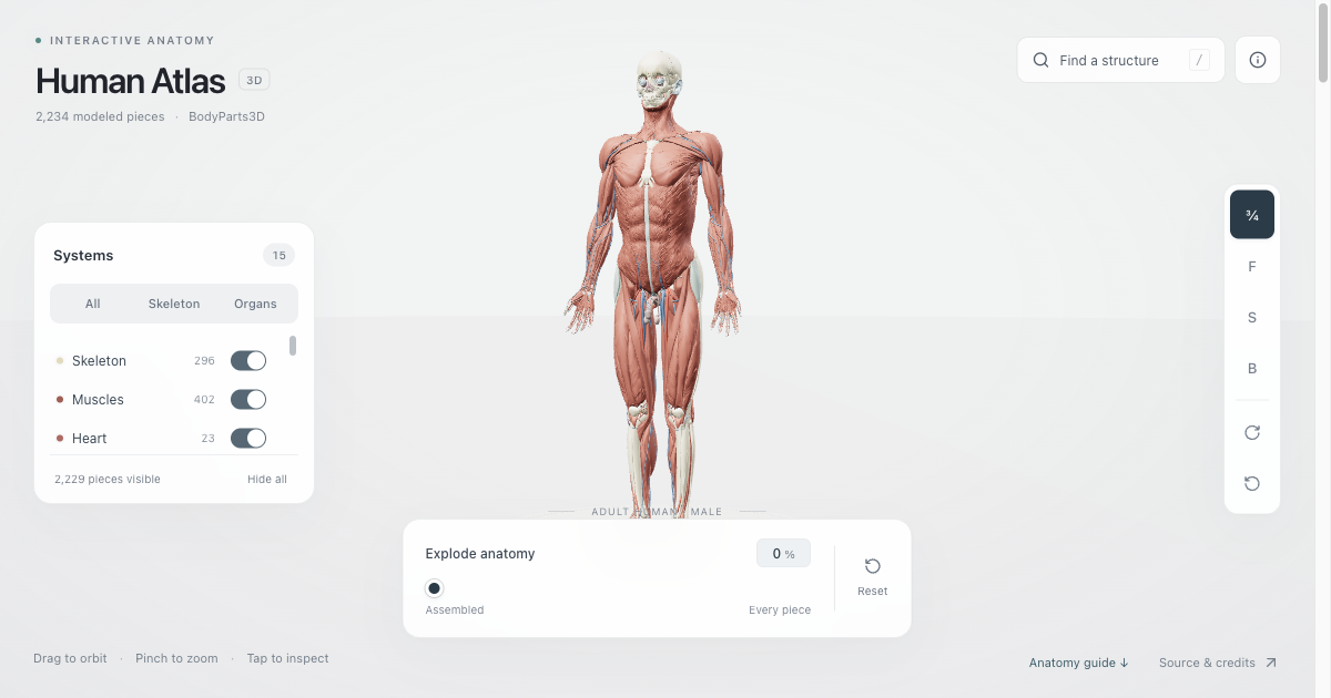
01 / GETTING STARTED
How to use Human Atlas
Begin with the assembled view. Drag the body to orbit around it, and use your scroll wheel or a pinch gesture to zoom. Front, side, back, and three quarter controls provide repeatable viewing angles.
Reveal one layer at a time
The Systems panel controls which groups are visible. Turn muscles off to expose deeper structures, choose Skeleton for a bone focused view, or select Organs to reduce the surrounding detail. Clicking a system name shows that group alone.
Find and inspect a structure
Search Human Atlas with an English anatomical name such as heart, brain, liver, or femur. Choose a result to open its details and highlight its associated pieces. A named concept can include several pieces, so its selection count may exceed one.
Read a view without losing context
The colors distinguish display groups; they are not a photograph of living tissue. When several layers overlap, hide one and compare the result before zooming further. The visible piece counter gives immediate feedback about how much anatomy remains on screen.
In a fully exploded view, pieces are arranged for browsing rather than kept in their original anatomical positions. Return the slider to Assembled before studying spatial relationships. If an organ seems unfamiliar from one direction, restore the surrounding body and switch to a standard camera view.
Separate, isolate, and reset
Use the explosion slider to spread visible pieces apart. For a closer look, select Isolate structure in the details panel. A reset control lets you restore the starting view, layers, and assembled arrangement.
02 / INSIDE THE BODY
Explore the systems in Human Atlas
Skeleton and muscles
Use Human Atlas to compare bones with the muscles around them. Switch between isolated systems and combined layers to observe their relative positions. The model supports visual exploration of shape and location; it does not simulate movement or muscle contraction.
Heart and blood vessels
The viewer separates the heart, arteries, and veins into display groups. Hide other systems to reduce visual clutter, then rotate the model to inspect vessel paths. Search for a specific named structure when the full vascular view becomes difficult to follow.
Nervous and sensory structures
The nervous system view reveals modeled neural structures, while sensory organs have a separate visibility control. Search for the brain or a cranial nerve to narrow your selection. This view provides spatial context rather than a simulation of nerve signals.
Respiratory and digestive organs
Here, respiratory and digestive layers help you compare structures within the trunk. Try searching for the trachea, stomach, or liver. Inspect one organ first, then restore its surroundings to understand how its position relates to other visible structures.
Other anatomical groups
The explorer also includes urinary, lymphatic, endocrine, reproductive, body surface, and connective tissue groups. Their piece counts vary with the source model. This is a specific reference dataset, so a small display group does not imply that a system is biologically simple.
Combined views
Combine layers when you want to compare neighboring structures. Start with two systems, inspect the overlap, and add another only when useful. A clear view often teaches more about a relationship than a crowded view with every layer enabled.
03 / A SIMPLE STUDY ROUTINE
Build a focused Human Atlas study session
Give each Human Atlas session a small, concrete question: where is this structure, what lies beside it, or how does its outline change from another angle? A focused question makes the controls easier to use and your observations easier to explain.
- Locate. Search for one structure and read the displayed name. Check the selected piece count before assuming you are looking at a single mesh.
- Inspect. Isolate the selection, rotate slowly, and compare front and side views. Describe the visible shape in your own words.
- Reconnect. Restore surrounding anatomy and identify two nearby structures. Toggle a layer to see which relationships were previously obscured.
- Review. Reset the viewer and locate the same structure again. Compare your observations with an anatomy textbook, course material, or the linked source documentation.
Use Human Atlas alongside your existing learning resources. The short descriptions provide general educational context, and some selections show a system overview rather than a detailed explanation of that individual structure. The displayed source identifier helps distinguish similarly named concepts.
04 / COMMON QUESTIONS
Questions about Human Atlas
Is Human Atlas free to use?
Yes. The explorer opens in your browser without a subscription, login, or API key. The application code is open source, and the anatomy data has a separate attribution license. Source links below explain where the project and model data come from.
Does Human Atlas work on a phone?
The mobile layout includes compact touch controls. Tap Systems in the bottom dock to reveal layer choices, drag to rotate, and pinch to zoom. Rendering performance depends on your device and browser; the first visit needs to load the model before exploration is ready.
Why are there different counts for pieces and concepts?
The viewer renders each source mesh once, but named anatomical concepts can group several meshes. A search result can therefore select multiple pieces. The 2,234 model pieces and 3,432 named concepts describe different aspects of the same reference, not two alternative body models.
Does Human Atlas show every human structure?
No. This model uses an adult male reference from BodyParts3D. It does not cover every structure, individual variation, age, or sex. Missing geometry should not be interpreted as an anatomical conclusion. Use broader references when you need information beyond this model.
Can Human Atlas be used for medical decisions?
This is an educational visualization, not a diagnostic or surgical tool. Geometry is simplified for browser performance, and display colors are chosen for exploration. The model cannot interpret symptoms, evaluate a scan, or provide information about a particular person's anatomy.
05 / SOURCE & SCOPE
The reference behind Human Atlas
Human Atlas uses BodyParts3D 4.0 geometry and anatomical metadata from the Database Center for Life Science. The web adaptation simplifies geometry and assigns display colors and system groups. These presentation choices make the model easier to explore while preserving the distinction between source meshes and named concepts.
The Human Atlas application is distributed under the MIT license. BodyParts3D data is licensed under Creative Commons Attribution 4.0 International. If you reuse the model, preserve the original attribution and identify adaptations. Read the included attribution document for the dataset credits and processing notes.
Return to the interactive model